Limb Lengthening and Reconstruction
External fixateurs
External fixateurs are frames that are applied to the limb using pins or tensioned wires and they allow the correction of deformity or management of fractures and avoid internal placement of plates or screws.
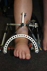
External fixateurs take a number of forms but the most common form these days is called a HEXAPOD.
A hexapod is two rings that are connected by six struts that can be adjusted in length with the aid of a web based software programme and this allows correction of all aspects of a deformity at the same time. Therefore a limb can be lengthened angulated translated and rotated all at the same time as required to correct a deformity.
The original external fixateur used for deformity correction was the ILIZAROV and this is still widely used around the world today and is highly versatile but the Hexapod can correct in one episode deformities that require one or more Ilizarov frames and that is why they are preferred over the Ilizarov.
My preferred hexapod is called the TL Hex manufactured by Orthofix.
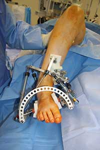
The TL Hex frame is applied under a general anaesthetic with fixation achieved with pins and wires as required. The shape of the frame matches the shape of the deformed limb and a cut in the bone(osteotomy) is performed.
Putting the information into a software programme allows the surgeon to provide the patient with a turning schedule and by following this “prescription” the patient corrects the deformity. The patient is seen regularly with clinical examination and X-rays to ensure it is progressing satisfactorily. At the end of adjustment the correction can be fine tuned with further turning schedules as required until the patient and surgeon are happy that the full correction has been achieved.
No further adjustments are carried out and the frame is removed once the bone has healed.
The TL Hex allows correction of complex deformity in a controlled and accurate manner that wouldn't be possible with standard internal fixation and acute correction techniques and is an extremely important part of our deformity correction “toolbox”.
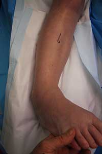
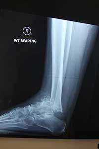
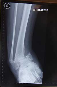
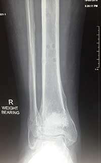
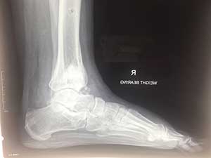
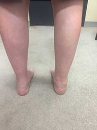
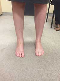
Pictures are a case study of a patient with a fracture complication from diabetes that had the external fixateur applied and the deformity gradually corrected with staged bone grafting- the fixateur was removed after three months with the fracture healed in an anatomical position
